Copyright
©2014 Baishideng Publishing Group Inc.
World J Gastrointest Pathophysiol. Aug 15, 2014; 5(3): 252-270
Published online Aug 15, 2014. doi: 10.4291/wjgp.v5.i3.252
Published online Aug 15, 2014. doi: 10.4291/wjgp.v5.i3.252
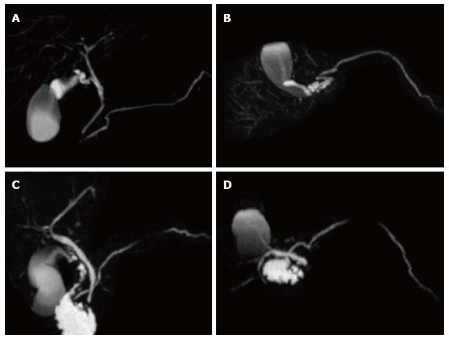
Figure 2 Normal pancreatic duct anatomy and pancreatic divisum.
(A and C) Coronal and (B and D) axial post-processed maximum intensity projection 3D-MRCP images from two different patients. In the first patient, the main pancreatic duct courses inferiorly (A) and posteriorly (B), joins the CBD and opens in the major papilla in keeping with normal pancreatic duct anatomy. In the second patient, the main pancreatic duct continues its course superiorly (C) and anteriorly (D), crosses the CBD and opens in the minor papilla in keeping with pancreatic divisum.
- Citation: Busireddy KK, AlObaidy M, Ramalho M, Kalubowila J, Baodong L, Santagostino I, Semelka RC. Pancreatitis-imaging approach. World J Gastrointest Pathophysiol 2014; 5(3): 252-270
- URL: https://www.wjgnet.com/2150-5330/full/v5/i3/252.htm
- DOI: https://dx.doi.org/10.4291/wjgp.v5.i3.252


