Copyright
©2013 Baishideng Publishing Group Co.
World J Gastrointest Pathophysiol. Nov 15, 2013; 4(4): 119-125
Published online Nov 15, 2013. doi: 10.4291/wjgp.v4.i4.119
Published online Nov 15, 2013. doi: 10.4291/wjgp.v4.i4.119
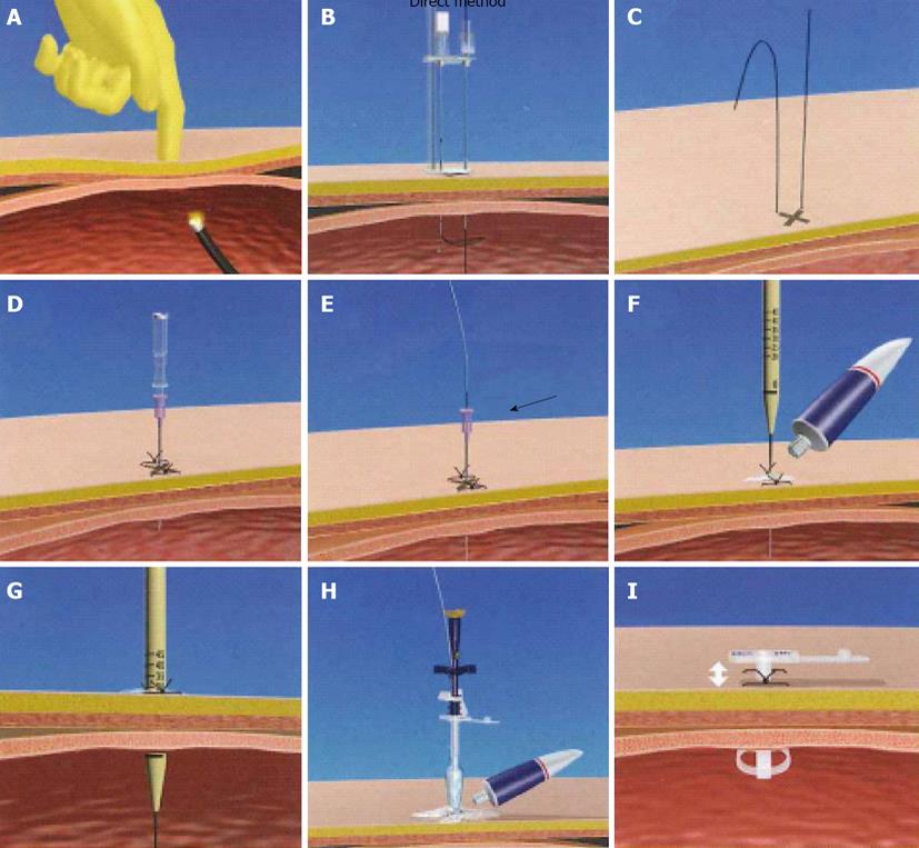
Figure 1 Direct method.
A: The transilluminated area on the abdominal wall was pushed with a finger; B, C: The stomach was punctured using a double-lumen gastropexy device; D: A needle with an outer plastic sheath (18-French) was introduced into the stomach under endoscopic control; E: The needle was removed and the guidewire was replaced; F, G: The skin incision was dilated by passing a dilator percutaneously into the stomach over the guidewire under endoscopic visualization; H: After the dilator was removed, a 24-French percutaneous endoscopic gastrostomy tube using an obturator was inserted over the guidewire; I: The tube was fixed to the abdominal wall.
- Citation: Ogino H, Akiho H. Usefulness of percutaneous endoscopic gastrostomy for supportive therapy of advanced aerodigestive cancer. World J Gastrointest Pathophysiol 2013; 4(4): 119-125
- URL: https://www.wjgnet.com/2150-5330/full/v4/i4/119.htm
- DOI: https://dx.doi.org/10.4291/wjgp.v4.i4.119


