Copyright
©2012 Baishideng Publishing Group Co.
World J Gastrointest Pathophysiol. Apr 15, 2012; 3(2): 51-59
Published online Apr 15, 2012. doi: 10.4291/wjgp.v3.i2.51
Published online Apr 15, 2012. doi: 10.4291/wjgp.v3.i2.51
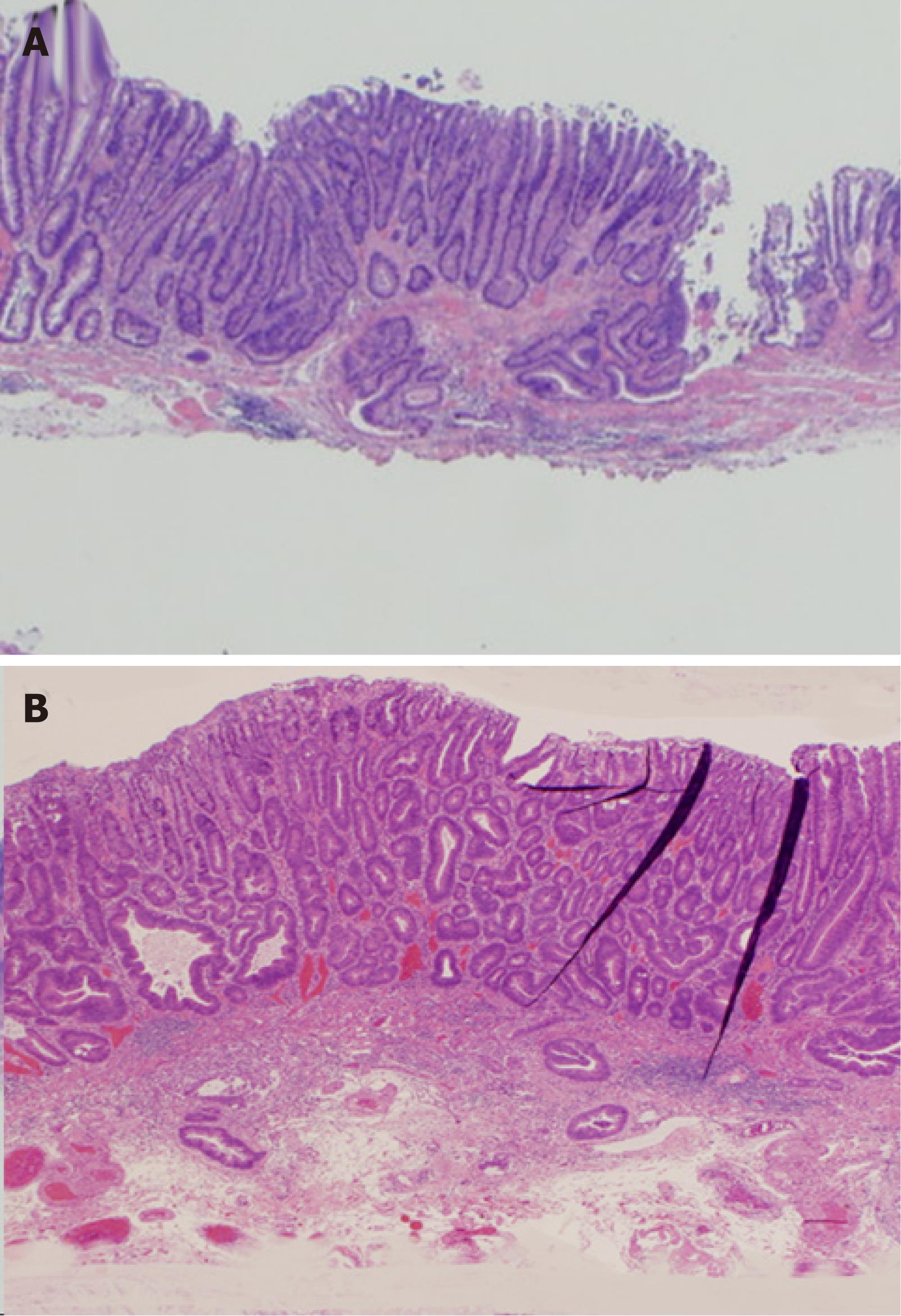
Figure 6 The depth of submucosal dissection in resection of submucosally invasive cancer by endoscopic submucosal dissection.
A: The dissection in this case was too shallow. Insufficient submucosa is seen in the resected specimen, which was dissected at the submucosa slightly below the muscularis mucosae. Submucosal invasion can be detected; however, the presence of venous-lymphatic invasion cannot be evaluated; B: This case was dissected appropriately. An adequate amount of submucosa is seen in the resected specimen, which was dissected at the middle-deep submucosa sufficiently below the muscularis mucosae. Both submucosal invasion and venous-lymphatic invasion can be detected.
- Citation: Yoshida N, Naito Y, Yagi N, Yanagisawa A. Importance of histological evaluation in endoscopic resection of early colorectal cancer. World J Gastrointest Pathophysiol 2012; 3(2): 51-59
- URL: https://www.wjgnet.com/2150-5330/full/v3/i2/51.htm
- DOI: https://dx.doi.org/10.4291/wjgp.v3.i2.51


