Copyright
©2012 Baishideng Publishing Group Co.
World J Gastrointest Pathophysiol. Apr 15, 2012; 3(2): 51-59
Published online Apr 15, 2012. doi: 10.4291/wjgp.v3.i2.51
Published online Apr 15, 2012. doi: 10.4291/wjgp.v3.i2.51
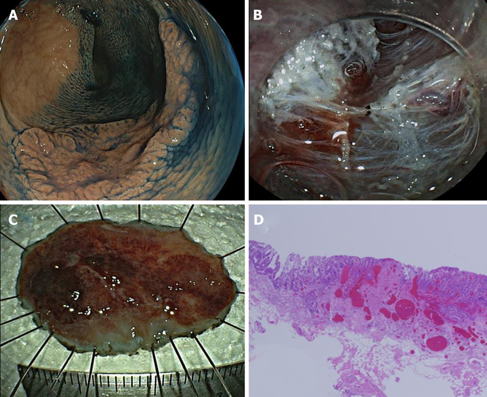
Figure 5 A submucosally invasive cancer with severe fibrosis.
A: A tumor graded 0-IIa, measuring 35 mm, located in the descending colon. The surface of the tumor was slightly depressed. The tumor was diagnosed by magnifying endoscopy as shallow submucosally invasive cancer and endoscopic submucosal dissection (ESD) was performed; B: Severe fibrosis was detected during ESD and was dissected with a scissor-type knife; C: En bloc resection was performed. The ESD operation time was 160 min. There was no perforation or postoperative hemorrhage; D: Histopathological diagnosis of the specimen resected by ESD was shallow submucosally invasive cancer. The depth of submucosal invasion was 800 μm, and there was severe fibrosis in the submucosa. ESD: Endoscopic submucosal dissection.
- Citation: Yoshida N, Naito Y, Yagi N, Yanagisawa A. Importance of histological evaluation in endoscopic resection of early colorectal cancer. World J Gastrointest Pathophysiol 2012; 3(2): 51-59
- URL: https://www.wjgnet.com/2150-5330/full/v3/i2/51.htm
- DOI: https://dx.doi.org/10.4291/wjgp.v3.i2.51


