Copyright
©2012 Baishideng Publishing Group Co.
World J Gastrointest Pathophysiol. Apr 15, 2012; 3(2): 44-50
Published online Apr 15, 2012. doi: 10.4291/wjgp.v3.i2.44
Published online Apr 15, 2012. doi: 10.4291/wjgp.v3.i2.44
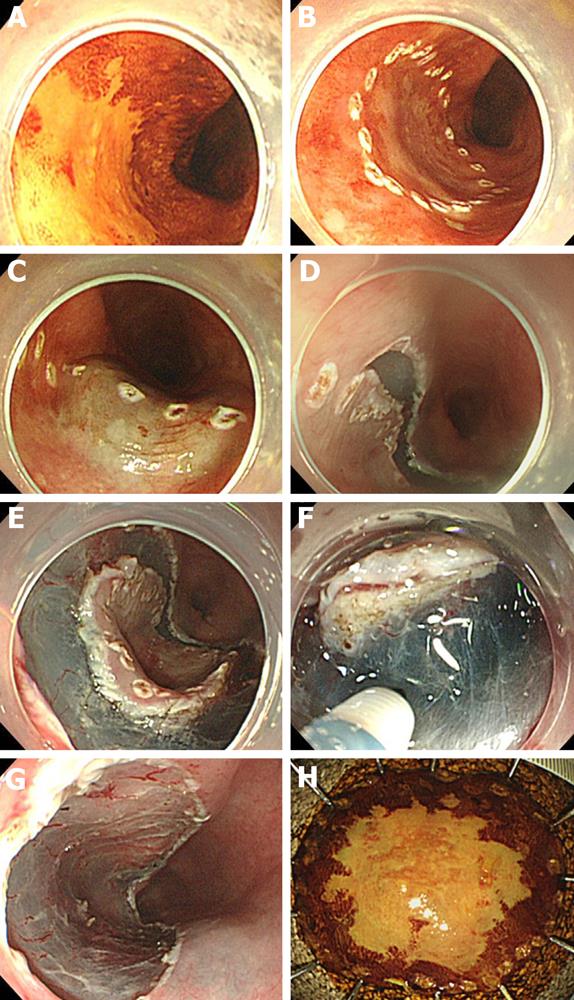
Figure 1 Steps of esophageal endoscopic submucosal dissection.
A: Iodine-unstained lesion revealed by chromoendoscopy with iodine staining; B: Marking around the lesion; C: Submucosal injection to generate a submucosal cushion; D: Mucosal incision around the marking dots from the distal side; E: Mucosal incision from the proximal side; F: Submucosal dissection beneath the lesion; G: Artificial ulcer after removal of the lesion; H: Resected en bloc specimen.
- Citation: Honda K, Akiho H. Endoscopic submucosal dissection for superficial esophageal squamous cell neoplasms. World J Gastrointest Pathophysiol 2012; 3(2): 44-50
- URL: https://www.wjgnet.com/2150-5330/full/v3/i2/44.htm
- DOI: https://dx.doi.org/10.4291/wjgp.v3.i2.44


