Copyright
©2012 Baishideng Publishing Group Co.
World J Gastrointest Pathophysiol. Feb 15, 2012; 3(1): 27-43
Published online Feb 15, 2012. doi: 10.4291/wjgp.v3.i1.27
Published online Feb 15, 2012. doi: 10.4291/wjgp.v3.i1.27
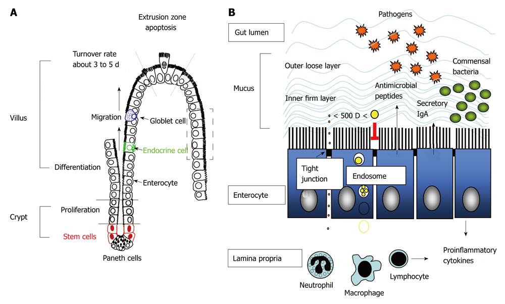
Figure 1 Intestinal crypt-villus axis and formation of intestinal barriers for luminal confinement of commensal bacteria.
A: Stem cells in the crypt regions undergo proliferation and differentiation into columnar epithelial cells (enterocytes) with high expression of brush border enzymes and transporters, and concurrently migrate upward to the apex of the villi where cell apoptosis and shedding occurs at the so-called “extrusion zone”. The stem cells also differentiate into Paneth cells that migrate downward to the crypt bottom, as well as into mucin-secreting goblet cells and enteroendocrine cells that migrate upwards to the villous tips. During the differentiation and migration process, tight junctional proteins are formed at the cell-cell contact sites to seal off gaps between enterocytes; B: Enteric microbes are restricted in the gut lumen by physical barriers composed of epithelium and mucus, chemical barriers with antimicrobial peptides, and immune barriers such as secretory immunoglobulin A (IgA). The tight junctional complexes between plasma membranes of two cells exclude the influx of bacteria and molecules larger than 500 dalton through paracellular routes, whereas endosomal degradation limits transcellular transport of particles and proteins. If the epithelial barrier is breached and invasion of bacteria occurs, the underlying immune cells in the lamina propria such as phagocytes (macrophages and neutrophils) and lymphocytes are responsible for antimicrobial and inflammatory responses.
- Citation: Yu LCH, Wang JT, Wei SC, Ni YH. Host-microbial interactions and regulation of intestinal epithelial barrier function: From physiology to pathology. World J Gastrointest Pathophysiol 2012; 3(1): 27-43
- URL: https://www.wjgnet.com/2150-5330/full/v3/i1/27.htm
- DOI: https://dx.doi.org/10.4291/wjgp.v3.i1.27


