Copyright
©2011 Baishideng Publishing Group Co.
World J Gastrointest Pathophysiol. Dec 15, 2011; 2(6): 88-92
Published online Dec 15, 2011. doi: 10.4291/wjgp.v2.i6.88
Published online Dec 15, 2011. doi: 10.4291/wjgp.v2.i6.88
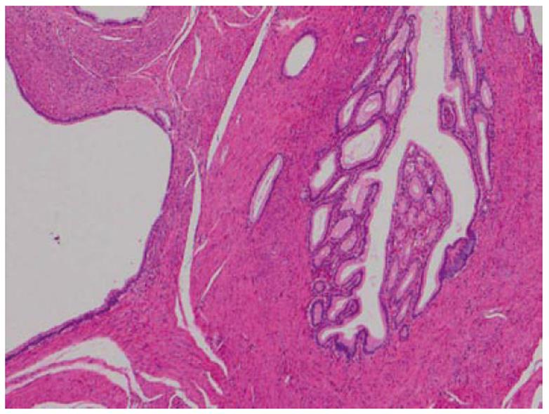
Figure 3 High-power view of adenomyoma of the small intestine.
The lesion consists of glandular structures of various sizes and interlacing smooth muscle bundles (hematoxylin and eosin stain, × 40).
- Citation: Takahashi Y, Fukusato T. Adenomyoma of the small intestine. World J Gastrointest Pathophysiol 2011; 2(6): 88-92
- URL: https://www.wjgnet.com/2150-5330/full/v2/i6/88.htm
- DOI: https://dx.doi.org/10.4291/wjgp.v2.i6.88


