Copyright
©2011 Baishideng Publishing Group Co.
World J Gastrointest Pathophysiol. Jun 15, 2011; 2(3): 53-56
Published online Jun 15, 2011. doi: 10.4291/wjgp.v2.i3.53
Published online Jun 15, 2011. doi: 10.4291/wjgp.v2.i3.53
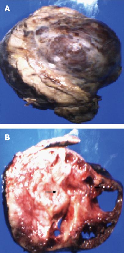
Figure 2 Gross photograph of the pancreatic tail tumor (A) and the corresponding cut surface (B) showing solid (thick arrow) and cystic areas (fine arrow)
- Citation: Bhavsar T, Wang C, Huang Y, Karachristos A, Inniss S. Littoral cell angiomas of the spleen associated with solid pseudopapillary tumor of the pancreas. World J Gastrointest Pathophysiol 2011; 2(3): 53-56
- URL: https://www.wjgnet.com/2150-5330/full/v2/i3/53.htm
- DOI: https://dx.doi.org/10.4291/wjgp.v2.i3.53


