Copyright
©The Author(s) 2020.
World J Gastrointest Pathophysiol. Jun 20, 2020; 11(4): 84-103
Published online Jun 20, 2020. doi: 10.4291/wjgp.v11.i4.84
Published online Jun 20, 2020. doi: 10.4291/wjgp.v11.i4.84
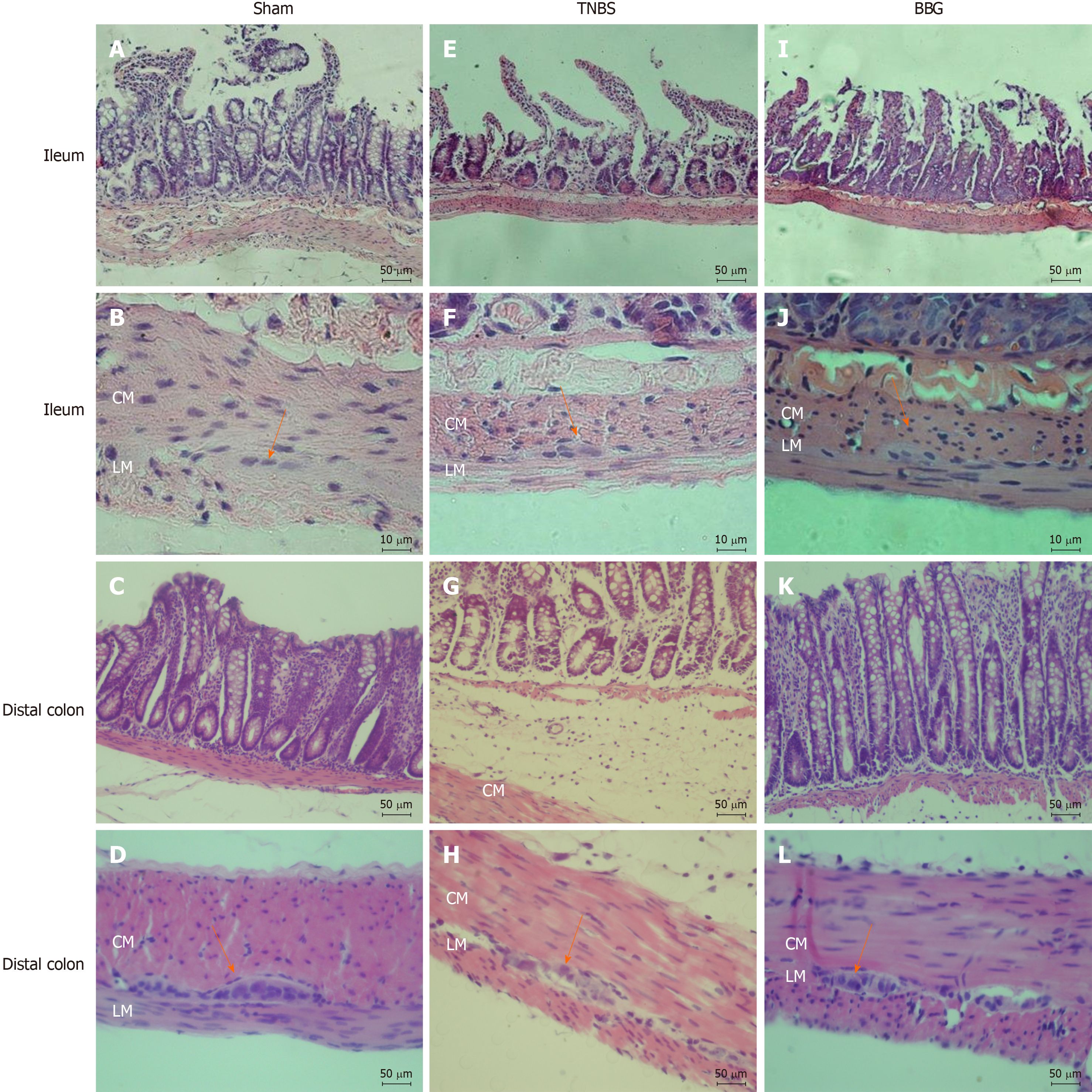
Figure 2 Photomicrographs showing sections stained with hematoxylin and eosin.
A, B, E, F, I, J: Rat ileum myenteric plexus in the sham, 2,4,6-trinitrobenzene sulfonic acid (TNBS) and brilliant blue G (BBG) groups; C, D, G, H, K, L: Rat distal colon myenteric plexus in the sham, TNBS and BBG groups. The histological observations showed that in ileum the appearances of the mucosa, circular and longitudinal muscles and enteric neurons in the sham, TNBS and BBG groups were preserved. However, the histological observations showed that in distal colon the edema and inflammatory cell infiltration in the TNBS group. The mucosa, the circular and longitudinal muscles and the distal colon enteric neurons in the sham and BBG groups were preserved. Orange arrows indicate myenteric ganglia. CM: Circular muscle; LM: Longitudinal plexus; TNBS: 2,4,6-trinitrobenzene sulfonic acid; BBG: Brilliant blue G. Scale bars: A, C, D, E, G, H, I, K, L = 50 μm; B, F, J = 10 μm.
- Citation: Souza RF, Evangelinellis MM, Mendes CE, Righetti M, Lourenço MCS, Castelucci P. P2X7 receptor antagonist recovers ileum myenteric neurons after experimental ulcerative colitis. World J Gastrointest Pathophysiol 2020; 11(4): 84-103
- URL: https://www.wjgnet.com/2150-5330/full/v11/i4/84.htm
- DOI: https://dx.doi.org/10.4291/wjgp.v11.i4.84


