Copyright
©The Author(s) 2020.
World J Gastrointest Pathophysiol. Jun 20, 2020; 11(4): 78-83
Published online Jun 20, 2020. doi: 10.4291/wjgp.v11.i4.78
Published online Jun 20, 2020. doi: 10.4291/wjgp.v11.i4.78
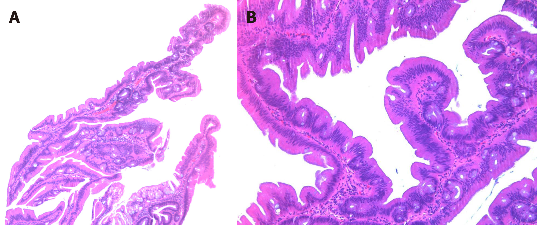
Figure 1 Low and high power view of the traditional serrated adenoma.
A: A low power view (40×) of the traditional serrated adenoma shows viliform growth of the polyp with slit-like serration; B: A high power view (100×) demonstrates ectopic crypt formation, eosinophilic cytoplasm and pencillate nuclei.
- Citation: Gui H, Husson MA, Mannan R. Correlations of morphology and molecular alterations in traditional serrated adenoma. World J Gastrointest Pathophysiol 2020; 11(4): 78-83
- URL: https://www.wjgnet.com/2150-5330/full/v11/i4/78.htm
- DOI: https://dx.doi.org/10.4291/wjgp.v11.i4.78


