Copyright
©2010 Baishideng Publishing Group Co.
World J Gastrointest Pathophysiol. Dec 15, 2010; 1(5): 154-165
Published online Dec 15, 2010. doi: 10.4291/wjgp.v1.i5.154
Published online Dec 15, 2010. doi: 10.4291/wjgp.v1.i5.154
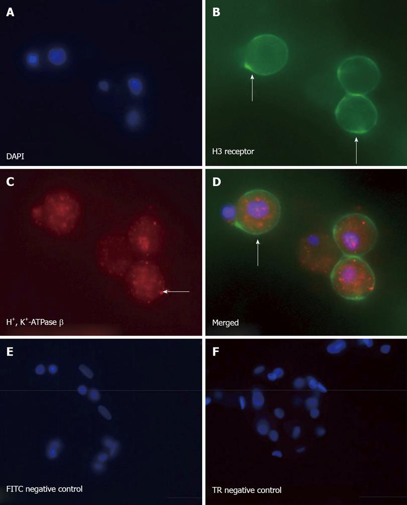
Figure 7 Primary canine parietal cells express the H3 receptor.
Immunofluorescence of isolated parietal cells stained with (A) 4', 6-Diamidino-2-Phenylindole
(DAPI), (B) H3 receptor (FITC), (C) H+, K+-ATPase b subunit (Texas red), and (D) merged image showing colocalization of the H3 receptor on the parietal cell. Negative IgG controls for (E) FITC and (F) Texas red (TR) secondary antibodies are shown.
-
Citation: Zavros Y, Mesiwala N, Waghray M, Todisco A, Shulkes A, Merchant JL. Histamine 3 receptor activation mediates inhibition of acid secretion during
Helicobacter -induced gastritis. World J Gastrointest Pathophysiol 2010; 1(5): 154-165 - URL: https://www.wjgnet.com/2150-5330/full/v1/i5/154.htm
- DOI: https://dx.doi.org/10.4291/wjgp.v1.i5.154


