Copyright
©The Author(s) 2017.
World J Radiol. Aug 28, 2017; 9(8): 330-338
Published online Aug 28, 2017. doi: 10.4329/wjr.v9.i8.330
Published online Aug 28, 2017. doi: 10.4329/wjr.v9.i8.330
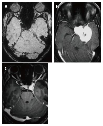
Figure 5 Contrast enhanced axial, susceptibility weighted imaging and 2 years follow up axial CE images.
A: SWI axial image shows isointense signal of the CSH; B: Contrast enhanced axial T1-W image demonstrates the postero-inferior extension of the CSH; C: Two years post-operative surveillance axial contrast enhanced T1-W image shows complete resolution of the lesion. T1-W: T1 weighted image; SWI: Susceptibility weighted image; CSH: Cavernous sinus hemangioma.
- Citation: Mahajan A, Rao VRK, Anantaram G, Polnaya AM, Desai S, Desai P, Vadapalli R, Panigrahi M. Clinical-radiological-pathological correlation of cavernous sinus hemangioma: Incremental value of diffusion-weighted imaging. World J Radiol 2017; 9(8): 330-338
- URL: https://www.wjgnet.com/1949-8470/full/v9/i8/330.htm
- DOI: https://dx.doi.org/10.4329/wjr.v9.i8.330


