Copyright
©The Author(s) 2017.
World J Radiol. Aug 28, 2017; 9(8): 330-338
Published online Aug 28, 2017. doi: 10.4329/wjr.v9.i8.330
Published online Aug 28, 2017. doi: 10.4329/wjr.v9.i8.330
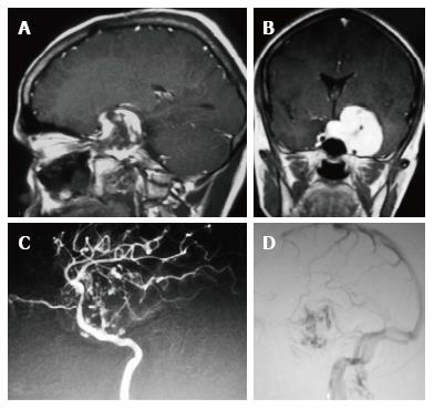
Figure 4 Contrast enhanced sagittal and coronal T1 weighted images and lateral views of digital subtraction angiography arterial and venous phases of left internal carotid angiogram.
A, B: Centripetal filling of the CSH in the left cavernous sinus is demonstrated; C: Arterial phase of DSA shows diffuse irregularity and stretching of C3 to C5 segments; D: Venous phase shows stasis in the venous channels within the CSH. CSH: Cavernous sinus hemangioma; DSA: Digital subtraction angiography.
- Citation: Mahajan A, Rao VRK, Anantaram G, Polnaya AM, Desai S, Desai P, Vadapalli R, Panigrahi M. Clinical-radiological-pathological correlation of cavernous sinus hemangioma: Incremental value of diffusion-weighted imaging. World J Radiol 2017; 9(8): 330-338
- URL: https://www.wjgnet.com/1949-8470/full/v9/i8/330.htm
- DOI: https://dx.doi.org/10.4329/wjr.v9.i8.330


