Copyright
©The Author(s) 2017.
World J Radiol. Aug 28, 2017; 9(8): 321-329
Published online Aug 28, 2017. doi: 10.4329/wjr.v9.i8.321
Published online Aug 28, 2017. doi: 10.4329/wjr.v9.i8.321
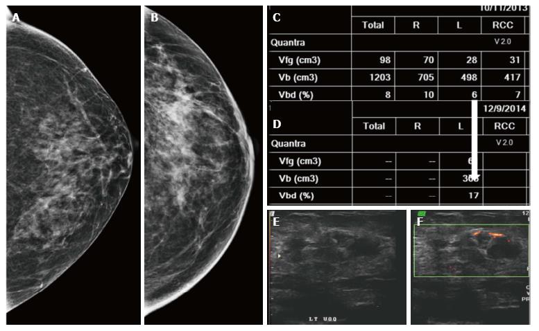
Figure 21 Patient underwent left breast-conserving therapy.
On follow-up mammograms at five (A) and six (B) years a subtle increase in density is noted. On volumetric assessment (C and D), the left breast density increased from 6 to 17; E and F: Ultrasound and color Doppler revealed a heterogeneously hypoechoic mass in left upper outer quadrant with peripheral vascularity. Histology: IDC.
- Citation: Ramani SK, Rastogi A, Mahajan A, Nair N, Shet T, Thakur MH. Imaging of the treated breast post breast conservation surgery/oncoplasty: Pictorial review. World J Radiol 2017; 9(8): 321-329
- URL: https://www.wjgnet.com/1949-8470/full/v9/i8/321.htm
- DOI: https://dx.doi.org/10.4329/wjr.v9.i8.321


