Copyright
©The Author(s) 2017.
World J Radiol. Aug 28, 2017; 9(8): 321-329
Published online Aug 28, 2017. doi: 10.4329/wjr.v9.i8.321
Published online Aug 28, 2017. doi: 10.4329/wjr.v9.i8.321
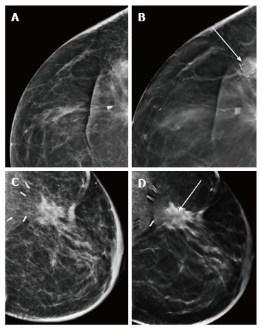
Figure 20 (A) and (B) represent conventional or 2D mammographic (A) and digital breast tomosynthesis (B) views of the scar site recurrence wherein mass density within the lesion is better appreciated on digital breast tomosynthesis (arrow); (C) and (D) show appearance of a scar on conventional or 2D mammogram (C) and digital breast tomosynthesis (D) where the fat lucency at the scar site is confirmed (arrow) on digital breast tomosynthesis.
- Citation: Ramani SK, Rastogi A, Mahajan A, Nair N, Shet T, Thakur MH. Imaging of the treated breast post breast conservation surgery/oncoplasty: Pictorial review. World J Radiol 2017; 9(8): 321-329
- URL: https://www.wjgnet.com/1949-8470/full/v9/i8/321.htm
- DOI: https://dx.doi.org/10.4329/wjr.v9.i8.321


