Copyright
©The Author(s) 2017.
World J Radiol. Aug 28, 2017; 9(8): 321-329
Published online Aug 28, 2017. doi: 10.4329/wjr.v9.i8.321
Published online Aug 28, 2017. doi: 10.4329/wjr.v9.i8.321
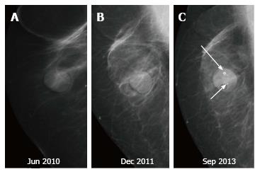
Figure 18 Microcalcifications in the nipple-areola region may be seen on the mammogram.
A: Three year post breast-conserving therapy mammogram - normal. Patient presented with unilateral yellowish nipple discharge and no clinically palpable abnormality at five year follow up. MMG revealed microcalcification (arrows in C) in the nipple-areola region. Histopathology: Paget’s disease of nipple; B: On retrospective evaluation, a tiny speck of calcification is seen in the nipple-areola region.
- Citation: Ramani SK, Rastogi A, Mahajan A, Nair N, Shet T, Thakur MH. Imaging of the treated breast post breast conservation surgery/oncoplasty: Pictorial review. World J Radiol 2017; 9(8): 321-329
- URL: https://www.wjgnet.com/1949-8470/full/v9/i8/321.htm
- DOI: https://dx.doi.org/10.4329/wjr.v9.i8.321


