Copyright
©The Author(s) 2017.
World J Radiol. Aug 28, 2017; 9(8): 321-329
Published online Aug 28, 2017. doi: 10.4329/wjr.v9.i8.321
Published online Aug 28, 2017. doi: 10.4329/wjr.v9.i8.321
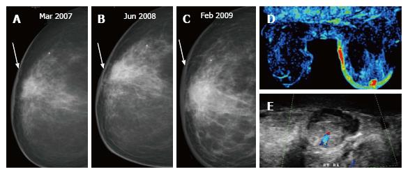
Figure 17 (A) In a post breast-conserving therapy patient, the skin thickness increases at one-year (B) and two-year (C) follow up mammograms; (D) dynamic MR perfusion reveals increased perfusion along the skin of the right breast and an enhancing focus, on ultrasound (E) an oval hypoechoic mass with increased vascularity is seen in the retroareolar region corresponding to the enhancing focus on breast MRI.
Histopathology: Angiosarcoma. MRI: Magnetic resonance imaging.
- Citation: Ramani SK, Rastogi A, Mahajan A, Nair N, Shet T, Thakur MH. Imaging of the treated breast post breast conservation surgery/oncoplasty: Pictorial review. World J Radiol 2017; 9(8): 321-329
- URL: https://www.wjgnet.com/1949-8470/full/v9/i8/321.htm
- DOI: https://dx.doi.org/10.4329/wjr.v9.i8.321


