Copyright
©The Author(s) 2017.
World J Radiol. Aug 28, 2017; 9(8): 321-329
Published online Aug 28, 2017. doi: 10.4329/wjr.v9.i8.321
Published online Aug 28, 2017. doi: 10.4329/wjr.v9.i8.321
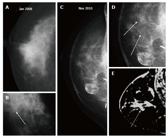
Figure 15 Microcalcifications.
A, B: Patient presented with a right breast mass with microcalcifications (arrow in B) and underwent right breast-conserving therapy with latissimus dorsi flap; C, D: At two years post treatment casting microcalcifications developed similar to the index lesion, better appreciated on magnified view (arrows in D) are seen around the scar site; E: Breast MRI revealed non-mass enhancement (dashed arrow) in the right breast. Histopathology of the recurrence showed DCIS. MRI: Magnetic resonance imaging.
- Citation: Ramani SK, Rastogi A, Mahajan A, Nair N, Shet T, Thakur MH. Imaging of the treated breast post breast conservation surgery/oncoplasty: Pictorial review. World J Radiol 2017; 9(8): 321-329
- URL: https://www.wjgnet.com/1949-8470/full/v9/i8/321.htm
- DOI: https://dx.doi.org/10.4329/wjr.v9.i8.321


