Copyright
©The Author(s) 2017.
World J Radiol. Aug 28, 2017; 9(8): 321-329
Published online Aug 28, 2017. doi: 10.4329/wjr.v9.i8.321
Published online Aug 28, 2017. doi: 10.4329/wjr.v9.i8.321
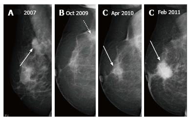
Figure 13 Patient with a right breast mass (arrow in A) who underwent breast-conserving therapy shows a normal post therapy mammogram at two years (B) with a post-surgical scar (dashed arrow), follow up imaging demonstrates a developing asymmetry in the retro-areolar region at three (C) years post therapy which subsequently developed into a frank mass (D).
- Citation: Ramani SK, Rastogi A, Mahajan A, Nair N, Shet T, Thakur MH. Imaging of the treated breast post breast conservation surgery/oncoplasty: Pictorial review. World J Radiol 2017; 9(8): 321-329
- URL: https://www.wjgnet.com/1949-8470/full/v9/i8/321.htm
- DOI: https://dx.doi.org/10.4329/wjr.v9.i8.321


