Copyright
©The Author(s) 2017.
World J Radiol. Aug 28, 2017; 9(8): 321-329
Published online Aug 28, 2017. doi: 10.4329/wjr.v9.i8.321
Published online Aug 28, 2017. doi: 10.4329/wjr.v9.i8.321
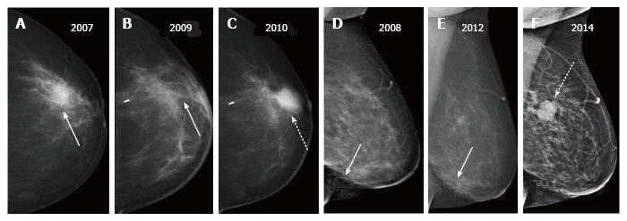
Figure 9 Two patients post breast-conserving therapy with recurrent masses.
A-C show a recurrent mass (dashed arrow in C) appearing at the scar site (solid arrow in A and B) two years after surgery; D-F demonstrate a post-surgical scar in the lower aspect (arrow in D and E) and a recurrent mass (dashed arrow in F) in upper aspect - different quadrant than the primary.
- Citation: Ramani SK, Rastogi A, Mahajan A, Nair N, Shet T, Thakur MH. Imaging of the treated breast post breast conservation surgery/oncoplasty: Pictorial review. World J Radiol 2017; 9(8): 321-329
- URL: https://www.wjgnet.com/1949-8470/full/v9/i8/321.htm
- DOI: https://dx.doi.org/10.4329/wjr.v9.i8.321


