Copyright
©The Author(s) 2017.
World J Radiol. Jul 28, 2017; 9(7): 304-311
Published online Jul 28, 2017. doi: 10.4329/wjr.v9.i7.304
Published online Jul 28, 2017. doi: 10.4329/wjr.v9.i7.304
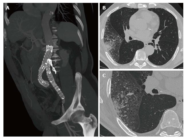
Figure 1 Lepidic predominant adenocarcinoma diagnosed during endovascular aortic aneurysm repair follow-up.
A-C: A 80-year-old male with a LPA of the right upper lobe diagnosed during endovascular aortic aneurysm repair follow-up for a type II endoleak treated with glue and coils (A). HRCT images (B and C) demonstrate the lepidic growth of the tumor and aerogenous metastases in the same lobe. LPA: Lepidic predominant adenocarcinoma; HRCT: High resolution computed tomography.
- Citation: Mazzei MA, Guerrini S, Gentili F, Galzerano G, Setacci F, Benevento D, Mazzei FG, Volterrani L, Setacci C. Incidental extravascular findings in computed tomographic angiography for planning or monitoring endovascular aortic aneurysm repair: Smoker patients, increased lung cancer prevalence? World J Radiol 2017; 9(7): 304-311
- URL: https://www.wjgnet.com/1949-8470/full/v9/i7/304.htm
- DOI: https://dx.doi.org/10.4329/wjr.v9.i7.304


