Copyright
©The Author(s) 2017.
World J Radiol. Jun 28, 2017; 9(6): 269-279
Published online Jun 28, 2017. doi: 10.4329/wjr.v9.i6.269
Published online Jun 28, 2017. doi: 10.4329/wjr.v9.i6.269
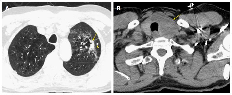
Figure 10 Challenging cases.
A: A 65-year-old man with a non-calcified spiculated lesion in the left upper lobe in the region of confluent pulmonary emphysema. The lesion reveals markedly irregular shape, with the solid component measuring 2.5 cm on computed tomography (CT) (arrow), which would put the lesion in the cT1c category. A separate solid nodule in the same lobe (arrowhead) would upstage the tumor to cT3; B: CT images at the cervicothoracic junction revealed large left thyroid lobe mass (arrow). The analysis of the left lung upper lobectomy specimen revealed a 3.2 cm primary lung adenocarcinoma, with the second nodule in the thyroid representing a metastatic thyroid cancer (pT2a lung cancer).
- Citation: Kay FU, Kandathil A, Batra K, Saboo SS, Abbara S, Rajiah P. Revisions to the Tumor, Node, Metastasis staging of lung cancer (8th edition): Rationale, radiologic findings and clinical implications. World J Radiol 2017; 9(6): 269-279
- URL: https://www.wjgnet.com/1949-8470/full/v9/i6/269.htm
- DOI: https://dx.doi.org/10.4329/wjr.v9.i6.269


