Copyright
©The Author(s) 2017.
World J Radiol. Jun 28, 2017; 9(6): 253-268
Published online Jun 28, 2017. doi: 10.4329/wjr.v9.i6.253
Published online Jun 28, 2017. doi: 10.4329/wjr.v9.i6.253
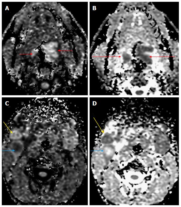
Figure 2 A 70-year-old male presented with a lesion in the base of tongue.
Diffusion weighted imaging revealed restricted diffusion (A) in the primary lesion (short arrows) with reduced ADC values (B, long arrows). Restricted diffusion was also noted in the two right level II nodes with reduced ADC values (C and D) indicating nodal metastases (yellow and blue arrows). One of the nodes, showed raised central ADC values suggestive of necrosis (blue arrow). Biopsy of the primary lesion revealed squamous cell carcinoma. ADC: Apparent diffusion coefficient.
- Citation: Mahajan A, Deshpande SS, Thakur MH. Diffusion magnetic resonance imaging: A molecular imaging tool caught between hope, hype and the real world of “personalized oncology”. World J Radiol 2017; 9(6): 253-268
- URL: https://www.wjgnet.com/1949-8470/full/v9/i6/253.htm
- DOI: https://dx.doi.org/10.4329/wjr.v9.i6.253


