Copyright
©The Author(s) 2017.
World J Radiol. May 28, 2017; 9(5): 230-244
Published online May 28, 2017. doi: 10.4329/wjr.v9.i5.230
Published online May 28, 2017. doi: 10.4329/wjr.v9.i5.230
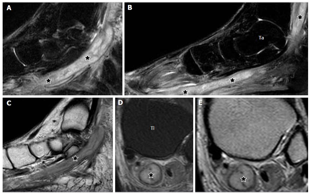
Figure 17 T2-weighted fat suppressed, serial sagittal (A, B); T1-weighted, sagittal (C); T2-weighted fat suppressed, axial (D) and proton density-weighted, axial (E) show an elongated tubular cystic lesion (stars) along the posterior tibial nerve at the lower leg, ankle and foot.
The lesion is seen within the substance of the nerve and has a central cystic component (stars) and a peripheral thin wall consistent with an abscess (D, E). This is a case of Hansen’s disease with posterior tibial nerve abscess; Ta: Talus; Ti: Tibia.
- Citation: Panwar J, Mathew A, Thomas BP. Cystic lesions of peripheral nerves: Are we missing the diagnosis of the intraneural ganglion cyst? World J Radiol 2017; 9(5): 230-244
- URL: https://www.wjgnet.com/1949-8470/full/v9/i5/230.htm
- DOI: https://dx.doi.org/10.4329/wjr.v9.i5.230


