Copyright
©The Author(s) 2017.
World J Radiol. May 28, 2017; 9(5): 230-244
Published online May 28, 2017. doi: 10.4329/wjr.v9.i5.230
Published online May 28, 2017. doi: 10.4329/wjr.v9.i5.230
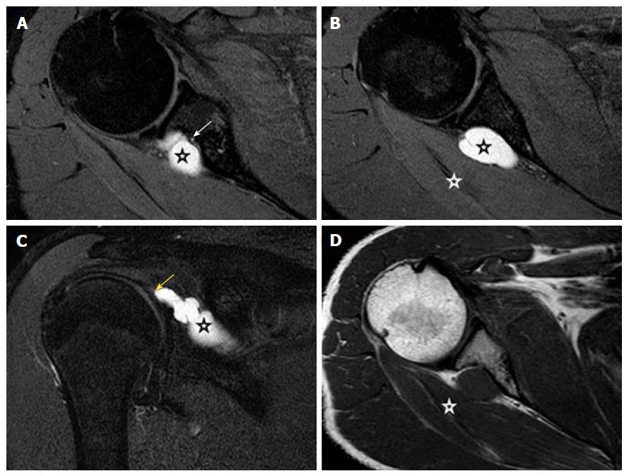
Figure 16 Proton density fat suppressed, serial axial (A, B), coronal (C) and T1-weighted, axial (D) show a well-defined lobulated slightly elongated cystic lesion (black stars) at the spinoglenoid notch compressing upon the suprascapular nerve (white arrow), which is seen separately from the cyst with preserved fat plane.
There is a tail like communication (yellow arrow) of the cyst with the posterior labrum. This suggests labral tear with paralabral cyst formation. Denervation edema and mild volume loss in the infraspinatus muscle (white stars) is seen.
- Citation: Panwar J, Mathew A, Thomas BP. Cystic lesions of peripheral nerves: Are we missing the diagnosis of the intraneural ganglion cyst? World J Radiol 2017; 9(5): 230-244
- URL: https://www.wjgnet.com/1949-8470/full/v9/i5/230.htm
- DOI: https://dx.doi.org/10.4329/wjr.v9.i5.230


