Copyright
©The Author(s) 2017.
World J Radiol. May 28, 2017; 9(5): 230-244
Published online May 28, 2017. doi: 10.4329/wjr.v9.i5.230
Published online May 28, 2017. doi: 10.4329/wjr.v9.i5.230
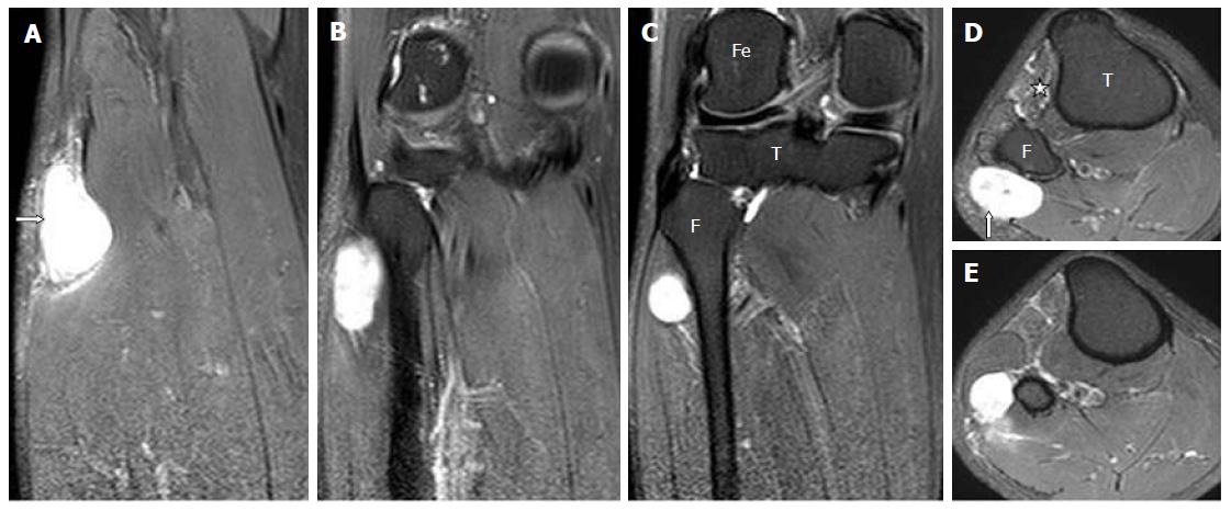
Figure 15 Serial, coronal (images A-C), and axial (images D-E), proton density fat suppressed sections show a well-defined oval cystic lesion at the posterolateral aspect of upper fibula along the expected course of common peroneal nerve which does not communicate within the proximal tibiofibular joint.
Mild denervation edema is seen in the anterior compartment muscles (star). This is a case of cystic schwannoma involving the CPN. T: Tibia; F: Fibula; Fe: Femur; CPN: Common peroneal nerve.
- Citation: Panwar J, Mathew A, Thomas BP. Cystic lesions of peripheral nerves: Are we missing the diagnosis of the intraneural ganglion cyst? World J Radiol 2017; 9(5): 230-244
- URL: https://www.wjgnet.com/1949-8470/full/v9/i5/230.htm
- DOI: https://dx.doi.org/10.4329/wjr.v9.i5.230


