Copyright
©The Author(s) 2017.
World J Radiol. May 28, 2017; 9(5): 230-244
Published online May 28, 2017. doi: 10.4329/wjr.v9.i5.230
Published online May 28, 2017. doi: 10.4329/wjr.v9.i5.230
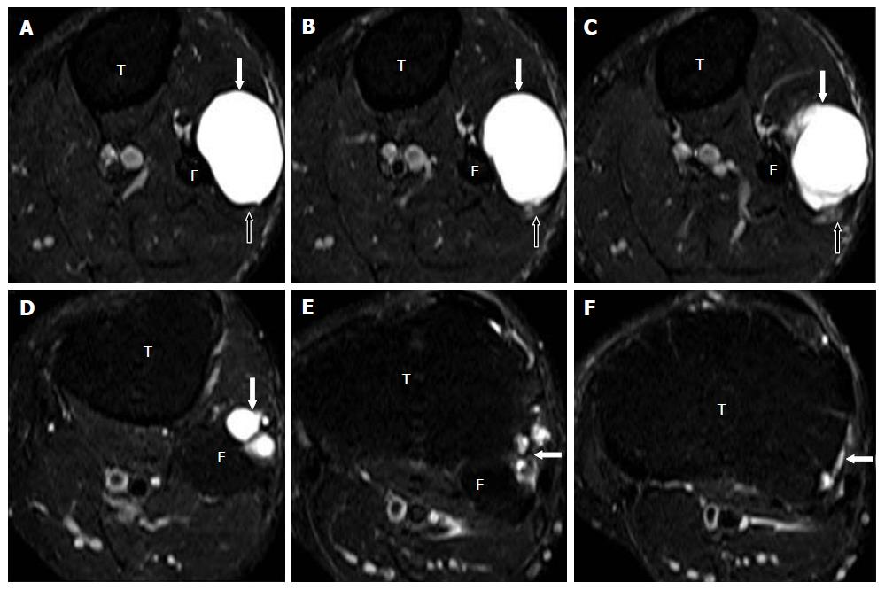
Figure 13 Images A-F show serial, T2-weighted fat suppressed axial sections of the proximal leg and demonstrate a large multilobulated globular extra-neural ganglion cyst (block arrows).
The ENGC is antero-lateral to the proximal fibula and indenting the peroneus longus muscle anteriorly (A-C). The CPN (open arrows) lies posterior to the cyst but is seen separate from it. The tail of the cyst (arrows in D-F) extends superiorly and communicates with the superior aspect of the PTF joint. PTF: Proximal tibiofibular; CPN: Common peroneal nerve; ENGC: Extraneural ganglion cyst; T: Tibia; F: Fibula.
- Citation: Panwar J, Mathew A, Thomas BP. Cystic lesions of peripheral nerves: Are we missing the diagnosis of the intraneural ganglion cyst? World J Radiol 2017; 9(5): 230-244
- URL: https://www.wjgnet.com/1949-8470/full/v9/i5/230.htm
- DOI: https://dx.doi.org/10.4329/wjr.v9.i5.230


