Copyright
©The Author(s) 2017.
World J Radiol. May 28, 2017; 9(5): 230-244
Published online May 28, 2017. doi: 10.4329/wjr.v9.i5.230
Published online May 28, 2017. doi: 10.4329/wjr.v9.i5.230
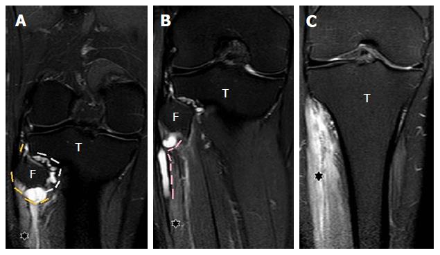
Figure 11 Images A-C show serial, coronal, proton density fat suppressed sections of the knee and proximal leg.
The entire extent of the cyst within the articular branch (white dashes) to the PTF joint extending to the CPN (yellow dashes) at the posterolateral fibular neck is seen, demonstrating the “u-sign” (A). The cyst extends into the proximal portion of the superficial peroneal nerve (pink dashes) for a length of approximately 5 cm (B). Denervation hyperintensity of the muscles (stars) of anterior and peroneal compartments of the leg is also seen. T: Tibia; F: Fibula; PTF: Proximal tibiofibular; CPN: Common peroneal nerve.
- Citation: Panwar J, Mathew A, Thomas BP. Cystic lesions of peripheral nerves: Are we missing the diagnosis of the intraneural ganglion cyst? World J Radiol 2017; 9(5): 230-244
- URL: https://www.wjgnet.com/1949-8470/full/v9/i5/230.htm
- DOI: https://dx.doi.org/10.4329/wjr.v9.i5.230


