Copyright
©The Author(s) 2017.
World J Radiol. May 28, 2017; 9(5): 230-244
Published online May 28, 2017. doi: 10.4329/wjr.v9.i5.230
Published online May 28, 2017. doi: 10.4329/wjr.v9.i5.230
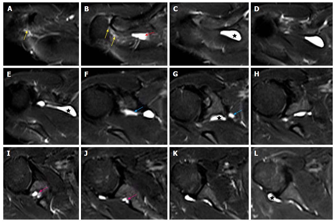
Figure 6 Images A-L show serial T2-weighted fat suppressed axial sections of the right shoulder, outlines the longitudinally oriented cyst (stars) along the course of the suprascapular nerve.
The cyst extends from the level of the acromioclavicular joint (B) to the posterior aspect of glenohumeral joint (L). A narrow joint connection extends along the expected course of the articular branch of the suprascapular nerve to the acromioclavicular joint (yellow arrows). Further descend of the intraneural cyst through the posterior triangle into the suprascapular and spinoglenoid notches are demonstrated by red, blue and pink arrows respectively. No obvious labral or capsular tear or degeneration of joint is noted on magnetic resonance imaging.
- Citation: Panwar J, Mathew A, Thomas BP. Cystic lesions of peripheral nerves: Are we missing the diagnosis of the intraneural ganglion cyst? World J Radiol 2017; 9(5): 230-244
- URL: https://www.wjgnet.com/1949-8470/full/v9/i5/230.htm
- DOI: https://dx.doi.org/10.4329/wjr.v9.i5.230


