Copyright
©The Author(s) 2017.
World J Radiol. May 28, 2017; 9(5): 230-244
Published online May 28, 2017. doi: 10.4329/wjr.v9.i5.230
Published online May 28, 2017. doi: 10.4329/wjr.v9.i5.230
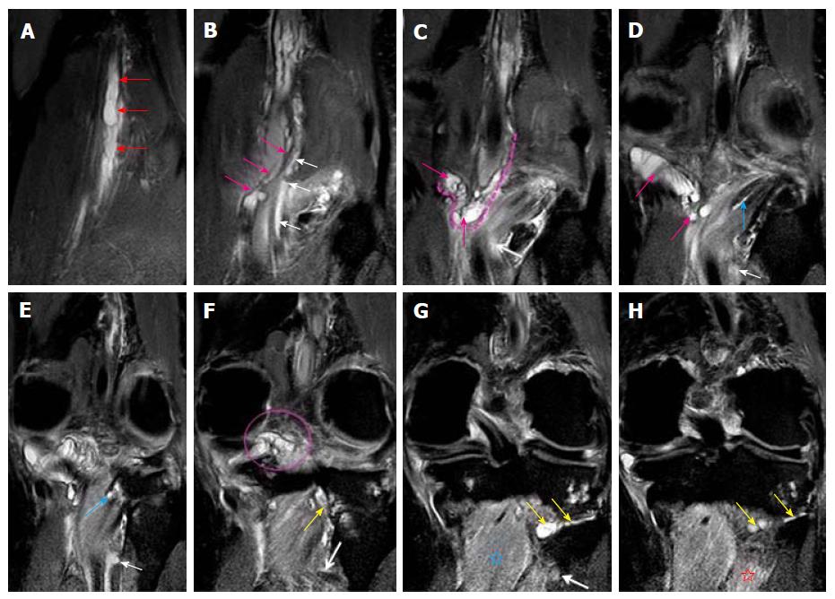
Figure 4 Images A-H show serial, proton density-weighted fat suppressed coronal sections of the knee demonstrate the longitudinal extent of intraneural cyst in the tibial nerve (red arrows), with propagation of cyst along the articular branches that communicate with the posterior aspect of knee joint (pink arrows, dashed line and circle) and to the postero-inferior part of proximal tibiofibular joint (yellow arrows).
This represents a dual joint connection (knee and proximal tibiofibular) from the same intraneural ganglion cyst. The cyst also extends along the branch to the popliteus (blue arrows) and tibialis posterior muscles (white arrows). Note the denervation edema in the popliteus (blue star) and tibialis posterior (red star) muscles. Superiorly, the cyst extends up to the bifurcation of the sciatic nerve in the distal third of the thigh.
- Citation: Panwar J, Mathew A, Thomas BP. Cystic lesions of peripheral nerves: Are we missing the diagnosis of the intraneural ganglion cyst? World J Radiol 2017; 9(5): 230-244
- URL: https://www.wjgnet.com/1949-8470/full/v9/i5/230.htm
- DOI: https://dx.doi.org/10.4329/wjr.v9.i5.230


