Copyright
©The Author(s) 2017.
World J Radiol. May 28, 2017; 9(5): 223-229
Published online May 28, 2017. doi: 10.4329/wjr.v9.i5.223
Published online May 28, 2017. doi: 10.4329/wjr.v9.i5.223
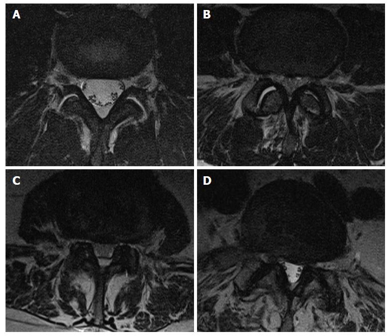
Figure 2 Axial T2-weighted magnetic resonance images illustrate the grading system of lateral recess stenosis.
A: Grade 0 bilaterally; B: Grade 1 bilaterally; C: Grade 2 bilaterally; D: Grade 3 on the left, Grade 1 on the right.
- Citation: Splettstößer A, Khan MF, Zimmermann B, Vogl TJ, Ackermann H, Middendorp M, Maataoui A. Correlation of lumbar lateral recess stenosis in magnetic resonance imaging and clinical symptoms. World J Radiol 2017; 9(5): 223-229
- URL: https://www.wjgnet.com/1949-8470/full/v9/i5/223.htm
- DOI: https://dx.doi.org/10.4329/wjr.v9.i5.223


