Copyright
©The Author(s) 2017.
World J Radiol. May 28, 2017; 9(5): 217-222
Published online May 28, 2017. doi: 10.4329/wjr.v9.i5.217
Published online May 28, 2017. doi: 10.4329/wjr.v9.i5.217
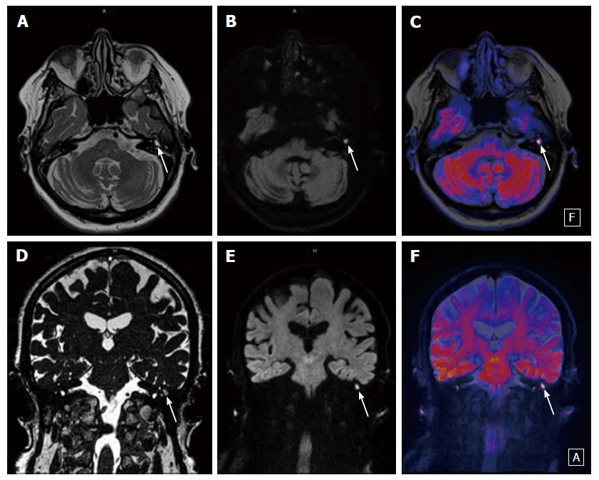
Figure 3 Twenty-three-year-old male patient with a typical cholesteatoma detected with diffusion weighted imaging.
A, D: T2 weighted images (A and D) show a fluid-like mass in the left middle ear (white arrow); B, C, E, F: EPI DWI RESOLVE (B and E) depict a hyperintense signal (white arrow) consistent with restriction due to a small cholesteatoma that is better demonstrated on fused images (C and F). DWI: Diffusion weighted imaging; EPI: Echo-planar imaging; RESOLVE: Readout-segmented echo-planar.
- Citation: Henninger B, Kremser C. Diffusion weighted imaging for the detection and evaluation of cholesteatoma. World J Radiol 2017; 9(5): 217-222
- URL: https://www.wjgnet.com/1949-8470/full/v9/i5/217.htm
- DOI: https://dx.doi.org/10.4329/wjr.v9.i5.217


