Copyright
©The Author(s) 2017.
World J Radiol. May 28, 2017; 9(5): 217-222
Published online May 28, 2017. doi: 10.4329/wjr.v9.i5.217
Published online May 28, 2017. doi: 10.4329/wjr.v9.i5.217
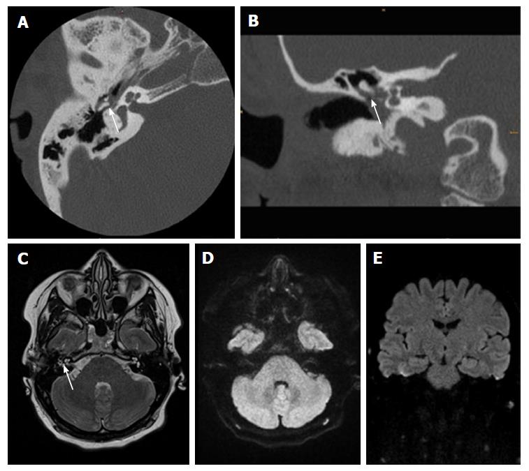
Figure 2 Thirty-one-year-old female after surgery for cholesteatoma.
A, B: CT show a soft-tissue mass in the tympanic space adjacent to malleolus and scutum with suspected bony erosion (white arrow); C-E: Axial T2 weighted image shows fluid-like signal (white arrow) that has no restriction in EPI DWI RESOLVE (axial in D and coronal in E). There was no sign of recurrent cholesteatoma on follow-up surgery. CT: Computed tomography; DWI: Diffusion weighted imaging; EPI: Echo-planar imaging; RESOLVE: Readout-segmented echo-planar.
- Citation: Henninger B, Kremser C. Diffusion weighted imaging for the detection and evaluation of cholesteatoma. World J Radiol 2017; 9(5): 217-222
- URL: https://www.wjgnet.com/1949-8470/full/v9/i5/217.htm
- DOI: https://dx.doi.org/10.4329/wjr.v9.i5.217


