Copyright
©The Author(s) 2017.
World J Radiol. Apr 28, 2017; 9(4): 191-198
Published online Apr 28, 2017. doi: 10.4329/wjr.v9.i4.191
Published online Apr 28, 2017. doi: 10.4329/wjr.v9.i4.191
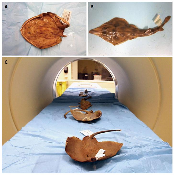
Figure 2 Formalin-preserved specimens positioned in the computed tomography scanner.
A: Skilletskate (Dactylobatus armatus); B: Southern Shovelnose ray (Aptychotrema vincentiana); C: The processes of the scan.
- Citation: McQuiston AD, Crawford C, Schoepf UJ, Varga-Szemes A, Canstein C, Renker M, De Cecco CN, Baumann S, Naylor GJP. Segmentations of the cartilaginous skeletons of chondrichthyan fishes by the use of state-of-the-art computed tomography. World J Radiol 2017; 9(4): 191-198
- URL: https://www.wjgnet.com/1949-8470/full/v9/i4/191.htm
- DOI: https://dx.doi.org/10.4329/wjr.v9.i4.191


