Copyright
©The Author(s) 2017.
World J Radiol. Apr 28, 2017; 9(4): 178-190
Published online Apr 28, 2017. doi: 10.4329/wjr.v9.i4.178
Published online Apr 28, 2017. doi: 10.4329/wjr.v9.i4.178
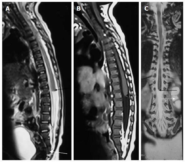
Figure 22 Caudal agenesis.
T2 weighted sagittal (A), T1 weighted sagittal (B) and T2 weighted coronal (C) MR images showing type II caudal agenesis. There is non-development of distal sacral vertebra (white arrow in A) with abnormal termination of conus medullaris (black arrow). The child also has associated left hydronephrosis (dashed arrow in C).
- Citation: Kumar J, Afsal M, Garg A. Imaging spectrum of spinal dysraphism on magnetic resonance: A pictorial review. World J Radiol 2017; 9(4): 178-190
- URL: https://www.wjgnet.com/1949-8470/full/v9/i4/178.htm
- DOI: https://dx.doi.org/10.4329/wjr.v9.i4.178


