Copyright
©The Author(s) 2017.
World J Radiol. Apr 28, 2017; 9(4): 178-190
Published online Apr 28, 2017. doi: 10.4329/wjr.v9.i4.178
Published online Apr 28, 2017. doi: 10.4329/wjr.v9.i4.178
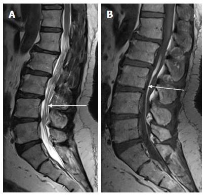
Figure 16 Fat in filumterminale.
T2 weighted (A) and T1 weighted (B) sagittal images of lumbosacral spine in a 60-year-old male showing linear T1 and T2 hyperintense focus in filumterminale (arrow) noted incidentally. Spinal cord is not low lying and there is no tethering of cauda equina fibres. Note is made of degenerative changes in lumbar spine.
- Citation: Kumar J, Afsal M, Garg A. Imaging spectrum of spinal dysraphism on magnetic resonance: A pictorial review. World J Radiol 2017; 9(4): 178-190
- URL: https://www.wjgnet.com/1949-8470/full/v9/i4/178.htm
- DOI: https://dx.doi.org/10.4329/wjr.v9.i4.178


