Copyright
©The Author(s) 2017.
World J Radiol. Apr 28, 2017; 9(4): 155-177
Published online Apr 28, 2017. doi: 10.4329/wjr.v9.i4.155
Published online Apr 28, 2017. doi: 10.4329/wjr.v9.i4.155
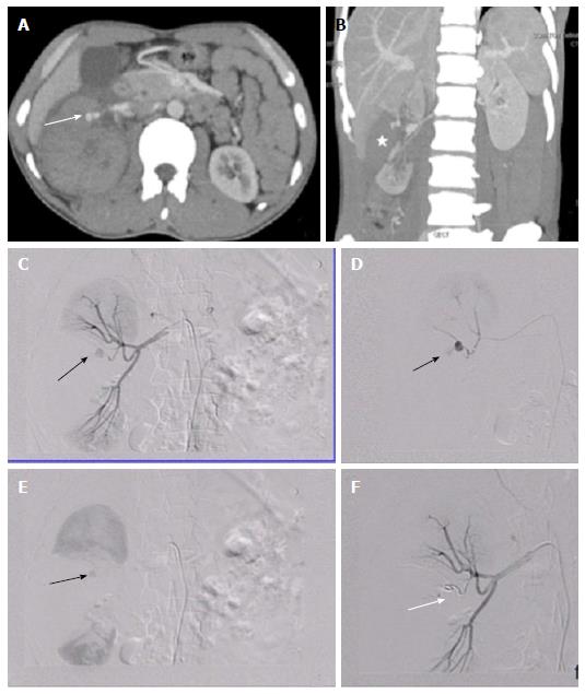
Figure 10 Renovascular injury: 35 years old male with blunt trauma abdomen.
A, B: CECT abdomen showed deep lacerations involving the interpole of right kidney extending upto the renal hilum (asterisk). A pseudoaneurysm (arrow) was seen arising from upper polar branch alongwith presence of perinephric hematoma; C, D: DSA with catheterisation of right main renal artery confirmed the presence of pseudoaneurysm (arrow); E: In view of end arterial supply, involved branch was selectively accessed and catheter was subsequently manipulated across the neck of PsA with the help of microwire followed by deployment of coil; F: Post embolization angiography showed non opacification distal to the coil (arrow). CECT: Contrast enhanced computed tomography; PsA: Pseudoaneurysm; DSA: Digital subtraction angiography.
- Citation: Singh A, Kumar A, Kumar P, Kumar S, Gamanagatti S. “Beyond saving lives”: Current perspectives of interventional radiology in trauma. World J Radiol 2017; 9(4): 155-177
- URL: https://www.wjgnet.com/1949-8470/full/v9/i4/155.htm
- DOI: https://dx.doi.org/10.4329/wjr.v9.i4.155


