Copyright
©The Author(s) 2017.
World J Radiol. Apr 28, 2017; 9(4): 155-177
Published online Apr 28, 2017. doi: 10.4329/wjr.v9.i4.155
Published online Apr 28, 2017. doi: 10.4329/wjr.v9.i4.155
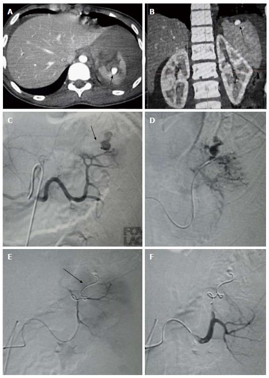
Figure 8 Splenic artery pseudoaneurysm.
Case of 39-year-old female with BTA, FAST positive, hemodynamically stable. CECT revealed (A and B) Extensive lacerations involving spleen with presence of contrast filled intrasplenic outpouching paralleling the aortic attenuation s/o pseudoaneurysm. Splenic artery angiogram showed (C) PsA (arrow) arising from the upper polar branch which on delayed phases showed contrast spillage s/o leaking PsA (D). Subsequently, upper pole branch was selectively catheterised by microcatheter followed by deployment of microcoils (arrow) across the neck of PsA (distal embolization) (E). Post embolization angiography (F) showed no flow distal to coil with exclusion of PsA and good contrast opacification of lower pole branch. CECT: Contrast enhanced computed tomography; BTA: Blunt trauma abdomen; PsA: Pseudoaneurysm; FAST: Focused assessment with sonography in trauma.
- Citation: Singh A, Kumar A, Kumar P, Kumar S, Gamanagatti S. “Beyond saving lives”: Current perspectives of interventional radiology in trauma. World J Radiol 2017; 9(4): 155-177
- URL: https://www.wjgnet.com/1949-8470/full/v9/i4/155.htm
- DOI: https://dx.doi.org/10.4329/wjr.v9.i4.155


