Copyright
©The Author(s) 2017.
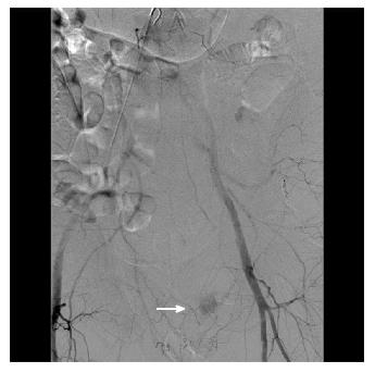
Figure 5 During interventional angiography, contrast extravasation is visualized into the colon via a distal branch artery from the internal iliac artery (white arrow).
The culprit vessel was occluded by spasm or dissection at the ostium with no residual active bleeding. During follow-up lower endoscopy, an endoscopic image showed no active bleeding with discrete large sized clean based ulcers, consistent with ischemic colitis.
- Citation: Ray DM, Srinivasan I, Tang SJ, Vilmann AS, Vilmann P, McCowan TC, Patel AM. Complementary roles of interventional radiology and therapeutic endoscopy in gastroenterology. World J Radiol 2017; 9(3): 97-111
- URL: https://www.wjgnet.com/1949-8470/full/v9/i3/97.htm
- DOI: https://dx.doi.org/10.4329/wjr.v9.i3.97


