Copyright
©The Author(s) 2017.
World J Radiol. Mar 28, 2017; 9(3): 126-133
Published online Mar 28, 2017. doi: 10.4329/wjr.v9.i3.126
Published online Mar 28, 2017. doi: 10.4329/wjr.v9.i3.126
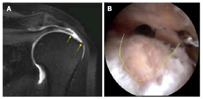
Figure 1 Magnetic resonance arthrography of a C2 lesion of the supraspinatus tendon.
A: MRA, coronal TSE T1w fat sat. Full tear with fiber retraction of supraspinatus tendon. Yellow arrows show the bare area of foot print lesion (C2 lesion according to Snyder classification); B: Arthroscopic view. Dotted line shows the crescent shape lesion. MRA: Magnetic resonance arthrography.
- Citation: Aliprandi A, Messina C, Arrigoni P, Bandirali M, Di Leo G, Longo S, Magnani S, Mattiuz C, Randelli F, Sdao S, Sardanelli F, Sconfienza LM, Randelli P. Reporting rotator cuff tears on magnetic resonance arthrography using the Snyder’s arthroscopic classification. World J Radiol 2017; 9(3): 126-133
- URL: https://www.wjgnet.com/1949-8470/full/v9/i3/126.htm
- DOI: https://dx.doi.org/10.4329/wjr.v9.i3.126


