Copyright
©The Author(s) 2017.
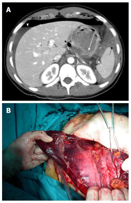
Figure 5 Gastric rupture.
A: Gastric wall rupture (white arrow): Peritoneal fluid (white arrowhead) with homogeneous hyperdense components from blood (black arrow), pneumoperitoneum with a bubble of gas located close to the gastric lesion (black arrowheads). Grade 4 lesion; B: Gastric rupture confirmed at surgery.
- Citation: Solazzo A, Lassandro G, Lassandro F. Gastric blunt traumatic injuries: A computed tomography grading classification. World J Radiol 2017; 9(2): 85-90
- URL: https://www.wjgnet.com/1949-8470/full/v9/i2/85.htm
- DOI: https://dx.doi.org/10.4329/wjr.v9.i2.85


