Copyright
©The Author(s) 2017.
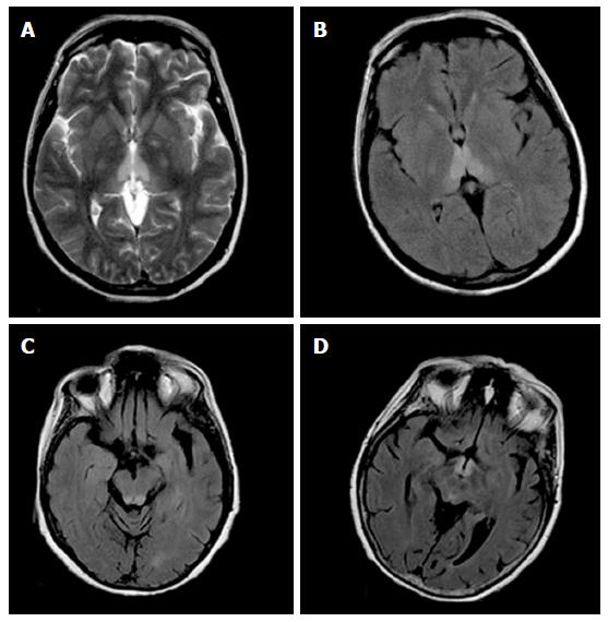
Figure 1 Magnetic resonance images in a patient with alcoholic Wernicke’s encephalopathy.
A: Axial T2-weighted image shows symmetrical high signal intensity lesions in the medial thalami; B: Axial fluid-attenuated inversion recovery (FLAIR) image shows symmetrical high signal intensity lesions in the medial thalami; C: Axial FLAIR image shows symmetrical high signal intensity lesions in the periaqueductal area; D: Axial FLAIR image shows symmetrical high signal intensity lesions around the mammillary bodies.
- Citation: Sparacia G, Anastasi A, Speciale C, Agnello F, Banco A. Magnetic resonance imaging in the assessment of brain involvement in alcoholic and nonalcoholic Wernicke’s encephalopathy. World J Radiol 2017; 9(2): 72-78
- URL: https://www.wjgnet.com/1949-8470/full/v9/i2/72.htm
- DOI: https://dx.doi.org/10.4329/wjr.v9.i2.72


