Copyright
©The Author(s) 2017.
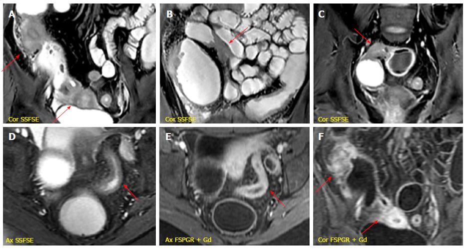
Figure 8 Fibrostenotic disease.
A-C: Multiple fibrotic strictures of the small bowel alternanting with prestenotic dilatated tracts detected on SSFSE sequences; D: Wall thickening of the sigmoid colon producing luminal narrowing displayed on SSFSE image; E and F: Post-gadolinium sequences reveal a diffuse and homogeneous enhancement in sigmoid colon (E) and small bowel (F) suggestive of subacute inflammation. SSFSE: Single shot fast spin echo; FSPGR: Fat-suppressed 3D spoiled gradient-echo.
- Citation: Mantarro A, Scalise P, Guidi E, Neri E. Magnetic resonance enterography in Crohn’s disease: How we do it and common imaging findings. World J Radiol 2017; 9(2): 46-54
- URL: https://www.wjgnet.com/1949-8470/full/v9/i2/46.htm
- DOI: https://dx.doi.org/10.4329/wjr.v9.i2.46


