Copyright
©The Author(s) 2017.
World J Radiol. Dec 28, 2017; 9(12): 438-447
Published online Dec 28, 2017. doi: 10.4329/wjr.v9.i12.438
Published online Dec 28, 2017. doi: 10.4329/wjr.v9.i12.438
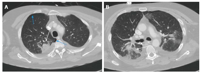
Figure 11 Computerised tomography scan of the chest 2 wk post-transplantation shows consolidation in the right upper lobe posteriorly with air bronchogram in keeping with pneumonia.
Associated right sided pneumothorax. A: Axial slice of CT chest showing right upper lobe consolidation (white arrow), and right sided pneumothorax (blue arrows); B: Axial slice showing consolidation on the right upper lobe with air bronchogram (arrow). CT: Computerised tomography.
- Citation: Chia E, Babawale SN. Imaging features of intrathoracic complications of lung transplantation: What the radiologists need to know. World J Radiol 2017; 9(12): 438-447
- URL: https://www.wjgnet.com/1949-8470/full/v9/i12/438.htm
- DOI: https://dx.doi.org/10.4329/wjr.v9.i12.438


