Copyright
©The Author(s) 2017.
World J Radiol. Oct 28, 2017; 9(10): 389-399
Published online Oct 28, 2017. doi: 10.4329/wjr.v9.i10.389
Published online Oct 28, 2017. doi: 10.4329/wjr.v9.i10.389
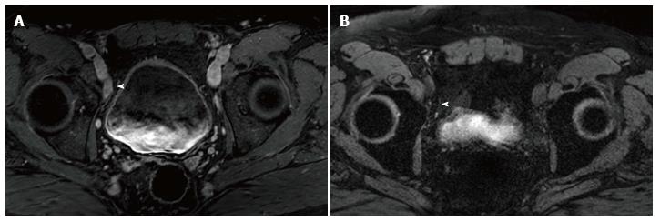
Figure 1 Selected imaged from a ferumoxytol enhanced magnetic resonance imaging in a 59-year-old man who underwent transrectal ultrasonography prostate biopsy for elevated PSA (10.
8 ng/mL) which showed Gleason 3 + 4 disease of 4 cores. A: Initial prostate multiparametric T1 weighted post gadolinium magnetic resonance imaging (MRI) showed a 1.8 cm × 0.9 cm right external iliac chain lymph node that was suspicious base on size criteria (arrowhead); B: 24 h post injection of ferumoxytol (7.5 Fe/kg dose), T2* weighted MRI showed decreased signal intensity within the node (arrowhead), consistent with uptake of ferumoxytol. This was considered a benign lymph node based on these results. The patient underwent computed tomography guided biopsy for confirmation and the node was negative for malignancy.
- Citation: Zarzour JG, Galgano S, McConathy J, Thomas JV, Rais-Bahrami S. Lymph node imaging in initial staging of prostate cancer: An overview and update. World J Radiol 2017; 9(10): 389-399
- URL: https://www.wjgnet.com/1949-8470/full/v9/i10/389.htm
- DOI: https://dx.doi.org/10.4329/wjr.v9.i10.389


