Copyright
©The Author(s) 2017.
World J Radiol. Oct 28, 2017; 9(10): 371-388
Published online Oct 28, 2017. doi: 10.4329/wjr.v9.i10.371
Published online Oct 28, 2017. doi: 10.4329/wjr.v9.i10.371
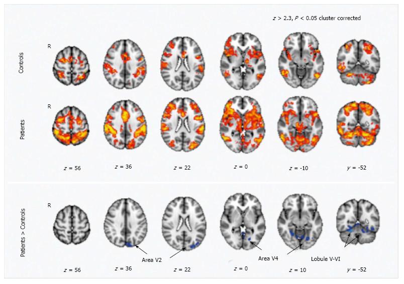
Figure 4 Motor training-dependent functional magnetic resonance imaging signal changes in healthy volunteers and in MS patients.
Maps of training-related functional magnetic resonance imaging signal changes are reported in healthy volunteers (indicated as controls) and in patients (Z > 2.3, P < 0.05, cluster corrected). Comparison between patients and controls show a higher signal reduction in the patients in regions corresponding to the secondary visual areas (V2 and V4) and in the cerebellum (lobule V-VI) (from Ref. [100]). V5: Visual cortex; R5: Right hemisphere; fMRI: Functional magnetic resonance imaging.
- Citation: Mormina E, Petracca M, Bommarito G, Piaggio N, Cocozza S, Inglese M. Cerebellum and neurodegenerative diseases: Beyond conventional magnetic resonance imaging. World J Radiol 2017; 9(10): 371-388
- URL: https://www.wjgnet.com/1949-8470/full/v9/i10/371.htm
- DOI: https://dx.doi.org/10.4329/wjr.v9.i10.371


