Copyright
©The Author(s) 2017.
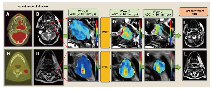
Figure 3 Representative no evidence of disease patient.
A and G: Pre-Tx PET/CT of the primary tumor and a neck nodal metastasis; B and H: Primary tumor and representative neck nodal metastasis contoured over a T2-W MRI; C and I: Pre-Tx ADC map overlaid on T2-W MRI; D and J: Wk2-Tx ADC map overlaid on T2-W; E and K: Wk3-Tx ADC map overlaid on T2-W; F and L: T2-W MRI post-Tx with no evidence of primary tumor and neck nodal metastases. PET/CT: Positron emission tomography/computed tomography; MRI: Magnetic resonance imaging; ADC: Apparent diffusion coefficient; IMRT: Intensity modulated radiation therapy.
- Citation: Aramburu Núñez D, Lopez Medina A, Mera Iglesias M, Salvador Gomez F, Dave A, Hatzoglou V, Paudyal R, Calzado A, Deasy JO, Shukla-Dave A, Muñoz VM. Multimodality functional imaging using DW-MRI and 18F-FDG-PET/CT during radiation therapy for human papillomavirus negative head and neck squamous cell carcinoma: Meixoeiro Hospital of Vigo Experience. World J Radiol 2017; 9(1): 17-26
- URL: https://www.wjgnet.com/1949-8470/full/v9/i1/17.htm
- DOI: https://dx.doi.org/10.4329/wjr.v9.i1.17


