Copyright
©The Author(s) 2017.
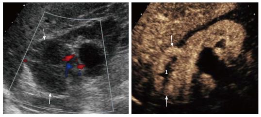
Figure 3 Patient with grade III chronic renal failure.
A: Color Doppler ultrasound showed markedly reduced renal parenchyma perfusion and a hypoechoic lesion without obvious vascularity (arrows); B: Contrast enhanced ultrasound showed a solid enhancing mass (arrows) with avascular central portion (1). A clear cell RCC with necrotic central areas was found at surgery. RCC: Renal cell carcinoma.
- Citation: Girometti R, Stocca T, Serena E, Granata A, Bertolotto M. Impact of contrast-enhanced ultrasound in patients with renal function impairment. World J Radiol 2017; 9(1): 10-16
- URL: https://www.wjgnet.com/1949-8470/full/v9/i1/10.htm
- DOI: https://dx.doi.org/10.4329/wjr.v9.i1.10


