Copyright
©The Author(s) 2016.
World J Radiol. Sep 28, 2016; 8(9): 785-798
Published online Sep 28, 2016. doi: 10.4329/wjr.v8.i9.785
Published online Sep 28, 2016. doi: 10.4329/wjr.v8.i9.785
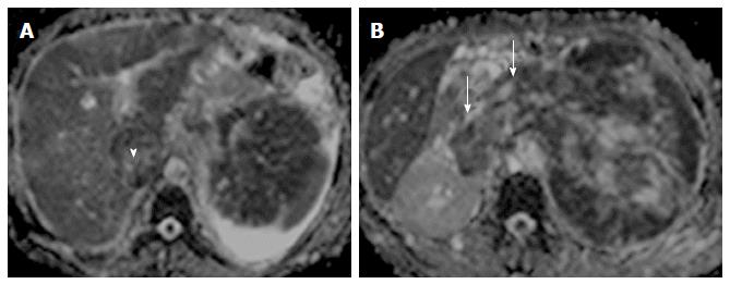
Figure 9 Renal cell carcinoma with malignant inferior vena cava thrombus.
Apparent diffusion coefficient maps (A and B) of a patient with large left renal mass with contiguous extension into renal vein (arrows) and inferior vena cava (arrowhead) (malignant thrombosis). Both the renal mass and intravascular thrombus had similar apparent diffusion coefficient values.
- Citation: Baliyan V, Das CJ, Sharma R, Gupta AK. Diffusion weighted imaging: Technique and applications. World J Radiol 2016; 8(9): 785-798
- URL: https://www.wjgnet.com/1949-8470/full/v8/i9/785.htm
- DOI: https://dx.doi.org/10.4329/wjr.v8.i9.785


