Copyright
©The Author(s) 2016.
World J Radiol. Sep 28, 2016; 8(9): 764-774
Published online Sep 28, 2016. doi: 10.4329/wjr.v8.i9.764
Published online Sep 28, 2016. doi: 10.4329/wjr.v8.i9.764
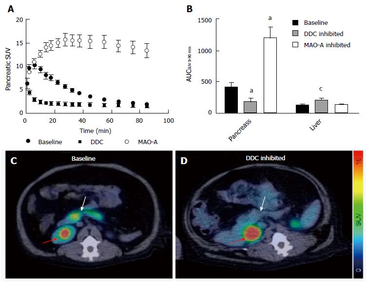
Figure 8 Positron emission tomography/computed tomography imaging of 5-hydroxy-L-11C-tryptophan in non-human primates.
A: SUV of [11C]5-HTP uptake in pancreas of nonhuman primate after 0-100 min p.i.; B: Pancreas uptake was significantly decreased by pretreated inhibitors (aP < 0.05; cP < 0.001); C and D: Abdominal HTP and PET/CT fusion images. Pancreas: White arrow; Kidney pelvis: Red arrow. Reprinted with permission from Ref.[65]. [11C]5-HTTP: 5-hydroxy-L-11C-tryptophan; PET/CT: Positron emission tomography/computed tomography. DDC:Dopa decarboxylase.
- Citation: Li J, Karunananthan J, Pelham B, Kandeel F. Imaging pancreatic islet cells by positron emission tomography. World J Radiol 2016; 8(9): 764-774
- URL: https://www.wjgnet.com/1949-8470/full/v8/i9/764.htm
- DOI: https://dx.doi.org/10.4329/wjr.v8.i9.764


